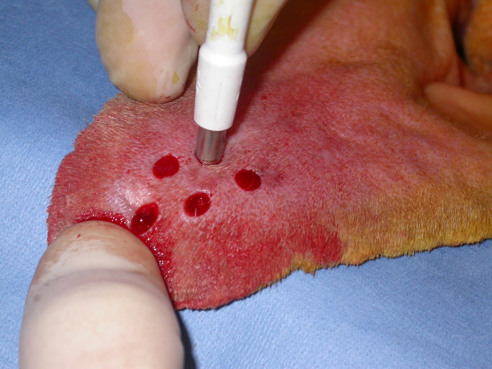

This study describes the relative popularity and perceived success of treatments used for aural haematoma in the dog. Left untreated, an ear hematoma will eventually heal on its own, but the pet will experience discomfort for weeks. If it is left untreated and infected all the more by a dog’s scratching or shaking, it can take the form of a severe swelling with excessive bleeding and can also cause impaired loss of hearing. Cosmetic results with medical management were excellent and with surgical treatment were good. Aural hematoma in dogs is caused due to the rupture of blood vessels in the floppy areas of a dog’s ears. This can be done using a needle and syringe or an indwelling plastic drain (see below). Hematomas/seromas can occur in both dogs and cats. How do you drain a dogs ear hematoma If your dog has a large or painful aural haematoma, it will need draining. However, hematomas and seromas can also occur within the head or brain, within other organs of the body and even on the ear (i.e., aural hematoma).
#Aural hematoma in dogs skin
Recurrent haematoma was treated more commonly with surgery (67%) than that of the initial presentation. Subdermal hematomas/seromas form under the skin and are probably the most commonly type of hematoma or seroma. The most common reason to select a particular treatment was previous success (76%).
#Aural hematoma in dogs plus
Surgical procedures included linear incision with sutures alone (35%) or sutures plus stents (24%) and an S-shaped incision with sutures (23%). On initial presentation, treatments included needle drainage with local deposition of corticosteroids (43%), surgery (29%) and needle drainage without corticosteroids (16%). The V-Bonnet is an adjustable ear wrap for dogs that secures the ears taking the stress away from bandaging and the e-collar. Aural haematomas are blood collections affecting the concave surface of the pinna and are more commonly seen in dogs than in cats (Lanz & Wood. Totally 312 email addresses were invalid, 259 questionnaires were completed (12♵% response rate) and 251 were included in analysis. They blood in the pinna is often clotted. If left untreated, the dog’s ear may become permanently deformed due to the formation of scar tissue and/or infection. Ear hematomas do require prompt veterinary treatment.

They are a collection of blood under the skin in the pinna or ear flap. Your dog may have an aural hematoma, a pocket of blood that accumulates between the ear’s cartilage and the skin. Questions investigated treatment selection for initial and repeat presentations of aural haematoma in dogs and their opinion of treatment success to prevent recurrence and for good cosmesis. In dogs, aural haematomas are very similar. Totally 2386 emails were sent to veterinary surgeons and practices inviting them to complete an online survey. An aural (ear) haematoma is a collection of blood or serum, and some- times a blood clot, within the pinna or ear flap. To survey the current treatment techniques of aural haematomas in dogs and investigate veterinary opinion regarding treatment success.


 0 kommentar(er)
